


Causes of papillomas in the neck
There's an etiological reason why papillomas start growing in the neck or in any other area of the human body - human papillomavirus (HPV) infection, which is a member of the family Papovaviridae. There are more than 100 serotypes of this pathogenic agent, each of which is responsible for the appearance of a different clinical picture of the disease (papilloma, condyloma, warts - these terms are synonymous, different names are associated with the sites in a particular area).
The major routes of transmission are contact household and genitals (condyloma in the perianal region). The virus is only able to penetrate the skin in the presence of microdamages or open wounds, in other cases it is not able to cross the protective barrier of the skin.
Pathogen Information
- It has a high degree of spread regardless of sex (however, it often manifests itself more often in women than in men), age or region (according to some sources, 2/3 of the planet is infected with this virus).
- Contains double-stranded, twisted ring DNA capable of integrating into the human genome.
- Infection with some strains is associated with a high carcinogenic risk, especially in the case of permanent damage. Throat papillomas are caused by non-oncogenic strains of the virus.
- A virus goes through two main stages in the sharing process. In the first stage, it is in episomal (free) form, and during the same period, the main division of viral particle occurs. This phase is reversible (after treatment a prolonged remission occurs). In the second - integrative step, the virus is implanted in the cell's genome (the first step towards cell degeneration and the formation of a malignant neoplasm). The first step is transient and passes relatively quickly, while the second is latent and explains the existence of carriers.
- The basal layer of the epidermis where the virus replicates is affected. In the remaining layers, the pathogen may persist but not divide. Provided that the virus is in the germ layer as it grows, the normal differentiation of cells in all layers in this area is disturbed, especially at the level of the prickly layer.
- Tends to have prolonged asymptomatic transport in the body (from several months to a year). It is rarely possible to identify a particular moment of infection - this is why treatment begins during a period of intense clinical manifestations and not at the first vague signs.
- To prevent infection, bivalent and quadrivalent vaccines are used, which are particularly effective against the most oncogenic strains 16 and 18.
Predisposing factors
- Lack of hygiene. As the virus is able to maintain vital activity in the external environment for a long time, it is necessary to strictly adhere to the rules of personal hygiene when visiting public places (swimming pool, bathhouse, gym).
- Traumatic skin damage. In order for the virus to penetrate, micro cracks or scratches on the skin (eg caused by rubbing the neck with the collar of a shirt) are enough.
- Impaired immune system function. With immune deficiencies of any origin, favorable conditions for the development of infections arise. For example, frequent colds and infectious diseases lead to a weakening of the immune system and the appearance of papillomas on the skin.
- Self-infection by scratching the skin.
- Systematic lifestyle disorder (stress, lack of physical activity, poor diet). These factors affect the work of all metabolic processes in the body and lead to a decrease in the barrier function of the skin.
- Environmental factors affecting the decline in the body's defenses (hypothermia, excessive ultraviolet exposure).
External manifestations of the disease
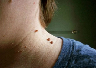
Cervical papillomas in the image look like this:
- The growth is most often located on a broad base and protrudes significantly beyond the skin surface. Less commonly, the base of the papilloma is represented by a thin bone (in this case, the formation takes a hanging position). In the second option, the risk of injury is much higher.
- The boundaries of education are smooth and clear.
- The color does not differ from the surrounding skin. In rare cases, it may be slightly lighter or darker than the adjacent tissues.
- The surface is often smooth, smooth. Sometimes growth is possible on top of the papilloma, making the surface ribbed.
- Diameter varies widely - from 1-3 mm to several centimeters (small diameter papillomas are more common).
- Position on any area of the neck (back, side front). Sometimes the face is involved.
As a rule, there are many lesions located along the skin folds.
In very rare cases, papillomas in the neck can become malignant, ie. degenerate into a skin tumor. This can occur as a result of infection with an oncogenic HPV strain.
Signs that may indicate a malignant transformation are as follows:
- color change and heterogeneity (polymorphism);
- boundary change (blur, loss of definition);
- the appearance of asymmetry (when drawing a line through the conditional center of the formation, two equal halves cannot be obtained);
- intensive growth;
- bleeding or ulceration (a non-specific symptom, as it is also characteristic of a simple neoplasm injury);
- itching, burning, peeling
- apostasy is formed (small daughter formations around the central).
The appearance of such signs does not necessarily mean degeneration of the papilloma, but it does mean that you should consult a doctor and undergo a differentiated diagnosis to find out if we are talking about a common inflamed mole or skin cancer.
How to get rid of papillomas on the neck
Treatment of papillomas in the neck is carried out only in a complex way with a simultaneous effect on the pathological focus on the skin and on the pathogen itself in the blood.
There are several ways to fight:
Method |
Description |
Medicine |
Using cytostatics, immune modulators are designed to suppress the replication of the viral agent in the affected area and reduce its concentration in the blood. Some drugs (keratolytics) are applied topically directly to destroy the growth of the skin (cauterize and cause tissue necrosis). |
Physical Methods |
Cryodestruction, laser therapy, electrocoagulation. They aim to get rid of papillomas on both the neck and other parts of the body. These methods allow you to restore the aesthetic appearance of open areas and remove the viral reservoir - the neoplasms of the skin itself, but they do not completely remove the virus from the body. |
Combination Therapy |
Combines the two previous options and is therefore most effective. |
Treatment of papillomas with folk remedies (eg celandine juice) is ineffective and often dangerous. In any case, a prerequisite is consultation with a doctor.
Physical methods of destruction
It is possible to effectively reduce formations using the following physical methods:
Method |
Description |
Local action with concentrated acid solutions |
A 1. 5% solution of zinc chloropropionate in 50% 2-chloropropionic acid, a combination of nitric acid, acetic acid, oxalic acid, lactic acid and copper nitrate trihydrate, etc. is used. in accordance with surgical rules. . . Apply the agent point by point with a spatula until the color of the formation changes to a lighter one (as soon as this has happened, further application should be stopped immediately). To cure papilloma, you need to perform 1-2 treatments on average. |
Electrocoagulation |
Using a special electric knife, a point cut is made of formations without affecting the underlying tissue (there is a minimal effect on healthy skin cells). The method is most convenient when the formation has a long stem and a small size. |
Cryodestruction |
Focus is exposed to liquid nitrogen, ultra-low temperature leads to tissue necrosis. It is good to clean this way of teaching with a broad base. The nitrogen action time is chosen by a specialist (1-5 minutes). After moxibustion, a burn forms which heals on average 10 days. |
Laser Removal |
The most modern and delicate approach to removing growth in prominent places such as the neck. Has the most positive reviews. Using a light guide from 5 sec to 3 minutes in continuous mode, they act on focus. The healing period is much shorter than with other methods (5-7 days). The technique is associated with minimal trauma to the surrounding tissues due to the high precision of the action. |
Classical surgical removal (scalpel excision) |
Used extremely rarely, only with large lesions or with suspected malignancy. The reason is that the lesions are often multiple, scattered around the neck and too small for excision. In addition, after surgical excision, scars may remain, which in itself creates a cosmetic defect. |